


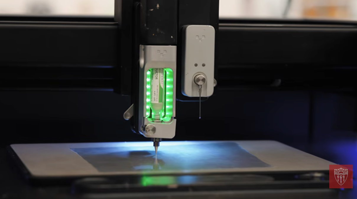
University of Southern California (USC) researchers have developed flexible, 3D printed MRI sensors that can be custom-made for individual infants and children in under 10 minutes for about $30, producing roughly four times the image contrast of standard commercial coils in testing.
Solving a Fit Problem for Small Patients
MRI machines rely on coils that act as antennas, picking up signals generated by the body’s response to magnets and radio waves. The tighter a coil fits around the area being scanned, the clearer the resulting image. Most coils, however, are built in standard adult sizes, and a poor fit around a small or rapidly growing body can weaken the signal and blur the image. Custom equipment has traditionally been the fix, but it can cost thousands of dollars and take months or years to produce, impractical for patients whose bodies change shape within weeks.
Yasser Khan, assistant professor of electrical and computer engineering and biomedical engineering at the USC Viterbi School of Engineering, led the effort out of his lab at the USC Michelson Center for Convergent Bioscience. “By making customized MRI equipment faster and more affordable to produce, we have the potential to bring better imaging to patients who have traditionally had fewer options, especially infants and children,” Khan said.
From Silver Ink to Skin-Like Material
Khan’s team spent about three years developing a manufacturing process that 3D prints conductive silver ink directly onto a thermoplastic elastomer, a soft, stretchable material with mechanical properties similar to human skin. The finished coil can stretch roughly 5% to 10%, letting it flex and move with the patient’s body instead of sitting rigidly against it. Getting there required testing multiple flexible materials, plastics, and printable metal formulations before settling on the silver-ink-and-elastomer combination, along with building custom electronics to connect the coils to existing MRI hardware.
Because each coil starts as a digital file, its dimensions can be adjusted on a computer and reprinted almost immediately, eliminating the wait for a manufacturer to fabricate a new physical part. “For a growing child, that could mean creating different coils as the body changes,” Khan said, adding that the goal is imaging equipment “designed for their bodies, so doctors can get the clearest picture possible as they grow.”
Paired with specialized imaging technology at the Michelson Center, the sensors have also shown potential for capturing fast-moving anatomy, including a beating heart.
Built on Cross-Disciplinary Collaboration
The project combined two labs’ expertise: Khan’s work on flexible and wearable electronics, and the Dynamic Imaging Science Center (DISC), led by Krishna Shrinivas Nayak, which develops MRI methods for imaging the body in motion using what researchers describe as a one-of-a-kind MRI system. Clinical input came from John Wood, director of cardiovascular MRI at Children’s Hospital Los Angeles, whose experience imaging children’s hearts, including real-time fetal heart imaging, helped guide where the technology could have the most impact.
“We need environments where different ideas can collide,” Khan said. “This project wouldn’t have happened without access to the MRI and conversations with cardiologists, radiologists and imaging scientists.”
A Long Push Toward Devices Built for Small Bodies
USC’s sensors are the latest example of a pattern in pediatric medical device research where researchers redesign the fabrication process itself so devices can be built directly around a child’s anatomy.
Sensor-integrated pediatric devices built this way aren’t new to 3D printing research. The University of Lincoln developed a 3D printed, sensor-operated prosthetic arm for toddlers under two years old, built around 3D scans of a child’s forearm to avoid the time-consuming plaster-casting typically used for socket modeling. This is a similar approach to USC for replacing slow, generic manufacturing with fast, personalized digital fabrication.
A closer parallel appeared when University College London researchers built a small 3D printed sensor to test brain responses in newborns, designing a wearable device safe for a baby’s face and testing it on infants just days old. Like USC’s coils, the device depended on precision fabrication at a scale standard manufacturing methods couldn’t easily reach.
Together, these projects point to a steady shift: 3D printing is increasingly the tool researchers reach for whenever the patient is simply too small for existing equipment to fit.
3D Printing Industry is inviting speakers for its 2026 Additive Manufacturing Applications (AMA) series, covering Energy, Healthcare, Automotive and Mobility, Aerospace, Space and Defense, and Software. Each online event focuses on real production deployments, qualification, and supply chain integration. Practitioners interested in contributing can complete the call for speakers form here.
To stay up to date with the latest 3D printing news, don’t forget to subscribe to the 3D Printing Industry newsletter or follow us on LinkedIn.
Explore the full Future of 3D Printing and Executive Survey series from 3D Printing Industry, featuring perspectives from CEOs, engineers, and industry leaders on the industrialization of additive manufacturing, 3D printing industry trends 2026, qualification, supply chains, and additive manufacturing industry analysis.
Featured image shows USC Team 3D Prints Custom MRI Sensors. Photo via USC.
