


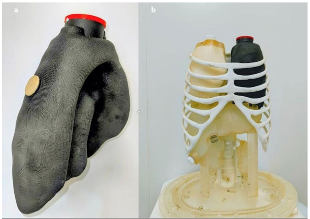
Researchers at University Hospital of Brest in France have 3D printed a fillable left lung model that simulates a pulmonary embolism (PE). They built it on a Formlabs Fuse 1+ selective laser sintering (SLS) system. The model is meant to give CT and nuclear medicine scans a known reference to measure against. In tests, a radiologist and two nuclear medicine physicians rated its images as highly realistic in all three imaging methods used.
The share of lung that loses blood flow after a clot, known as the pulmonary vascular obstruction index (PVOI), may help guide decisions on how long to continue anticoagulant treatment. PE is diagnosed mainly with CT and often followed up with nuclear medicine, but there is no independent ground truth for comparing how the two measure it.
Building a PE Lung From CT Data
The team scanned the healthy lung insert of a Kyoto Kagaku thorax phantom. They merged its contours with anonymized lung segmentation data in MIM Software’s image processing program. The superior segment of the left lower lobe became the embolism zone for three reasons:
- Its triangular shape resembles a typical perfusion defect.
- It makes up about 5% of lung volume, a commonly used PVOI cutoff.
- Its position at the back of the lung made it easier to fit a screw plug.
Two screw plugs added in Autodesk Fusion 360 let the healthy lung and the embolism compartment be filled separately. The researchers chose SLS because they considered it suited to producing a cavity nested inside a larger volume. The model had to be printed as a single block to stay watertight for radioactive solutions. The 14.3 × 24.1 × 10.4 cm part was printed in Nylon 12 at a 0.110 mm layer thickness and took 17 hours. It was printed off-site because the hospital has no large-format SLS printer.
How the Phantom Performed on Scans
The printed lung’s total volume came out 5.04% above the reference lung’s. Most of the difference was in the outer wall, which was 72% larger. The embolism compartment took up 7.6% of the internal volume, against a 5% design target. The team filled the model with polystyrene beads to reproduce lung tissue density. The authors say the measured densities were consistent with real lung, especially with 4 mm beads.
For testing, the healthy compartments were filled with:
- iodinated contrast for dual-energy CT on a GE Revolution HD
- 99mTc for planar scintigraphy on a Siemens system
- 68Ga for PET/CT on a Siemens system
The embolism segment was left empty each time. For the nuclear scans, the phantom sat on a moving platform linked to an Anzai respiratory gating system to simulate breathing. Two experienced readers, working independently and blinded, estimated the obstruction on the nuclear images at one segment, about 5% of lung volume. That result was consistent with the simulated defect.

Production Costs and Known Tradeoffs
Formlabs printed the phantom free of charge. The authors estimate that a team paying market rates would spend €770 on engineering, technician and printing time, nylon powder and shipping. That figure excludes the printer (€25,700) and its post-processing unit (€10,000), which outsourcing makes unnecessary.
The nylon walls are clearly visible on CT, so PVOI was not calculated from CT images. Only one lung segment was modeled. The simulated breathing ran along a single axis with a fixed lung volume, and some scans showed uneven bead and tracer distribution. The phantom is intended for future studies comparing how each imaging method measures perfusion defects, and for cross-scanner checks, quality control and protocol harmonization. The authors add that the workflow could be adapted to other organs, with organ-specific features redesigned each time.
Titled “Pulmonary embolism 3D printing model for inter-modality imaging study,” the study was conducted by Pauline Tripet, Mathieu Pavoine, Pierre-Yves Le Roux, Samuel Guigo, Brieg Dissaux, and David Bourhis.
Replacing Generic Phantoms with Task-Specific Prints
The study addresses a gap in physical references for PE imaging. Commercial torso phantoms can simulate lung imaging but generally have simplified, generic lung shapes that do not allow PE to be modeled realistically. The authors also say no commercially available phantom supported studies combining nuclear medicine with CT. Digital alternatives must be modeled and validated separately for each imaging method over several months. The authors say they found no earlier report of a fillable 3D printed lung phantom for PE assessment across both CT and nuclear medicine.
Stratasys and Siemens Healthineers start from a related premise. They introduced patient-specific 3D printed phantoms designed to enhance CT imaging, noting that current anatomical phantoms used in radiology lack the complexity needed for detailed diagnostic imaging. Like the Brest team, they use 3D printing to move beyond generic phantoms. Their models are built for CT and reproduce anatomy with precise radiopacity. The Brest model is a hollow shell filled with contrast or radioactive tracers so it can be read by both CT and nuclear medicine.
On the nuclear imaging side, Tran-Gia, Susanne Schlögl and Michael Lassmann 3D printed kidney phantoms in four sizes. The kidneys are waterproof and refillable, so they can be reused to set the amount of radioactive material needed for SPECT or CT imaging. Like the Brest lung, they are watertight, fillable organ shapes for radioactive solutions. The authors described the method as a promising prototyping technique for geometry-specific calibration of SPECT/CT systems. That calibration across organ sizes was their aim, while the Brest model is built to simulate a perfusion defect for comparing imaging methods.
Both examples use 3D printing to shape a phantom around a specific anatomy or imaging task, which is the role the Brest lung model is designed to fill for PE.
Vote now in the 2026 3D Printing Industry Awards.
To stay up to date with the latest 3D printing news, don’t forget to subscribe to the 3D Printing Industry newsletter or follow us on LinkedIn.
Explore the full Future of 3D Printing and Executive Survey series from 3D Printing Industry, featuring perspectives from CEOs, engineers, and industry leaders on the industrialization of additive manufacturing, 3D printing industry trends 2026, qualification, supply chains, and additive manufacturing industry analysis.
Featured image shows the insertion of the 3D printed lung prototype into the opened anthropomorphic phantom. Image via Tripet, et al., 3D Printing in Medicine.
